Vascular Wilts
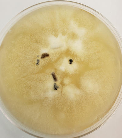
A culture of Ophiostoma growing on agar in a petri dish. A few pieces of elm wood are also on the plate.

The sporothrix stage producing conidia as seen in the lab. A better photo is below.
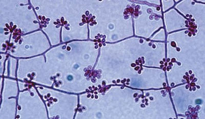
Sporothrix stage. Conidiophores producing conidia.
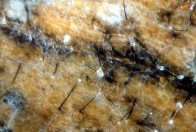
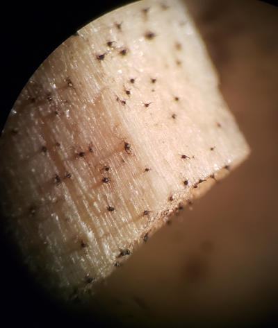
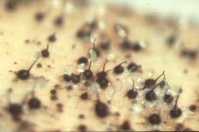
Synnemata on elm bark as viewed with a hand lens.

Another view of elm wood with synnemata of Ophiostoma. Conidia collect on the top of each synnema in a sticky droplet.
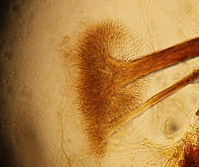
Microscopic view of the top of a synnema showing a flair of conidiophores that would produce the conidia.
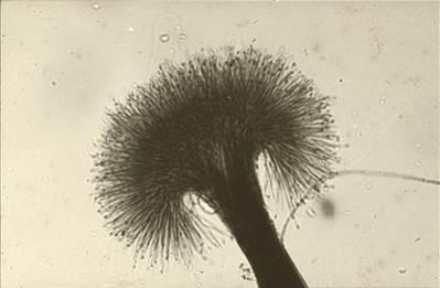
Another view of the top of the synemma showing many conidiophores producing very small conidia. The conidia accumulate in a sticky droplet on the synemma.
A and B strains coming together will result in perithecia forming on the elm wood.
Perithecia have a bulbous base and long neck. Ascospores accumulate on the tip in a sticky matrix.
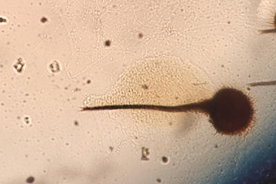
A perithecium as seen through the microscope with the spore mass oozing out.