Wood Decay Fungi Biology
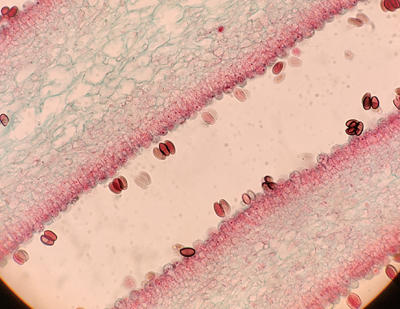
Section of a gill showing basidia and basidiospores on gill surfaces
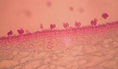
A higher magnification of a gill and basidiospores produced on basidia.
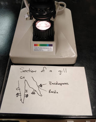
Diagram of the basdia and basidiospores on a gill.
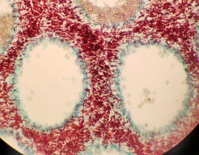
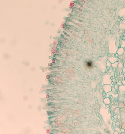
A section of a fruiting body with pores. The basidia (green), red is the mycelium between pores. The top left pore shows some basidiospores attached to basdidia.
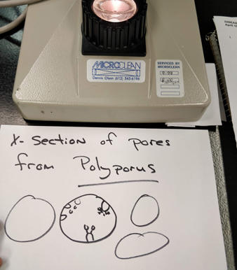
Diagram of basidia and basidiospores in pore.
A section of a fruiting body with teeth-like projections. The basidia and basidiospores line the surfaces of the "teeth". Some young immature basidiospores can be seen on the basidia. Red are detached mature basidiospores.
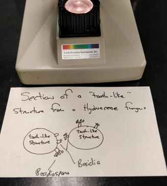
Basidia and basidospores produced on the surfaces of the teeth-like structures.
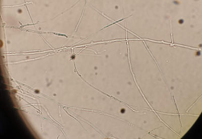
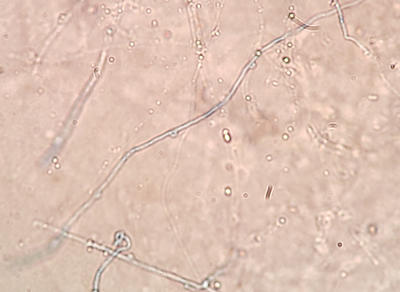
Mycelium of a wood decay fungus. Hyphae can be seen with clamp connections which look like small bumps on the hyphae.
Another wood decay fungus showing clamp connections on the hyphae seen in the middle of the photo.
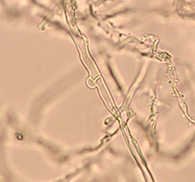
Another photo showing a higher magnification of a clamp connection from one of John's wood decay fungi he isolated from and mounted during the lab.
Brown rots
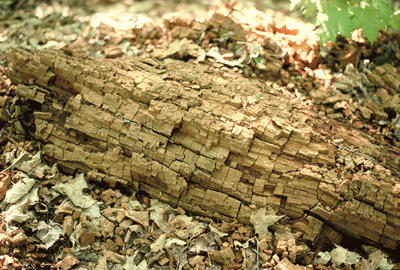
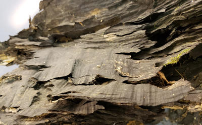
A fallen tree on the forest floor degraded by a brown rot fungus. The advanced decay often breaks apart into brown cubicle pieces.
Brown rot in aspen
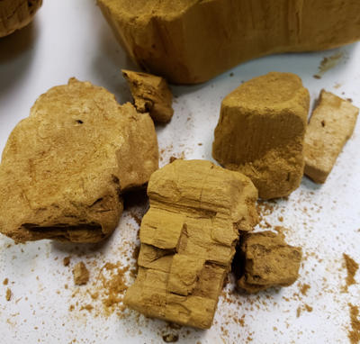
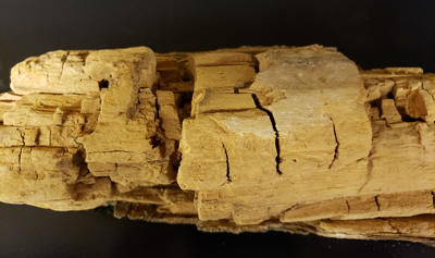
A closer look at the brown-rotted wood. When dry, it has little to no strength and breaks into fragments with slight pressure. When in the forest and wet, it can absorb moisture like a sponge.
White rots
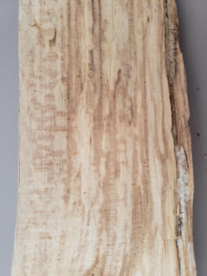
In the early stages of white rot, before substantial strength properties are lost, the wood becomes lighter in color.
White Rot in an aspen.
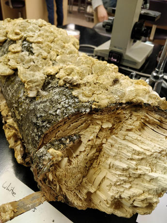
A birch tree with decay fungi fruiting and advanced stage of white rot.
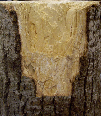
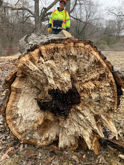
A white rot fungus in oak. When you have a fruiting body on a tree (upper right) it is usually associated with a lot of decay inside the tree.
Some white rot fungi are more selective at degrading lignin than others and cause a white pocket rot. The white "pockets" are pure cellulose and lignin has been completely removed.
Section of a large oak tree with very advanced white pocket rot caused by Inonotus dryophilus.
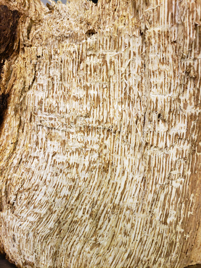
A close up of the white pocket rot in oak.
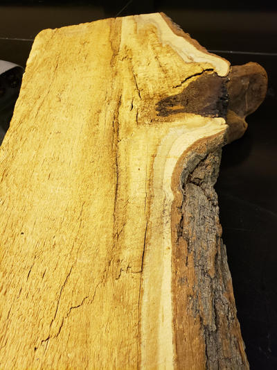
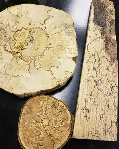
Zones lines are produced when incompatible white rot fungi meet. The pseudosclerotial plate is a three dimensional barrier that keeps other fungi from invading their territory. These barriers can also form near wood surfaces to keep out fungi and protect against moisture loss.
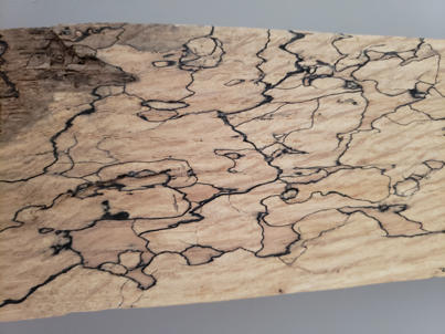
The zone lines produced interesting patterns in wood. In early stages of white rot, this wood can be used for wood working since not much wood strength loss or biomass lost has taken place.
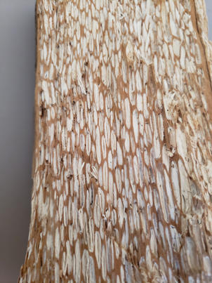
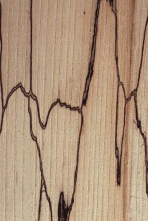
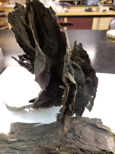
Some additional examples of zone lines produced by white rot fungi.
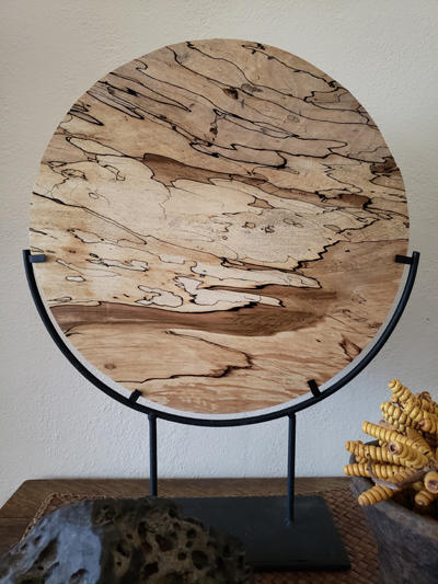
Some extraordinary art can be made from wood with zone lines. Here is a piece with many different populations of decay fungi producing many different zone lines as each tries to maintain their niche. Note also the different stages of white rot and wood coloration from brownish (or normal wood color), to very white.
The zone lines are made up of compounds that resist attack and degradation by other fungi and also insects. Over time, as the wood all gets degraded, the zone lines persist.
Another view of zones lines after all the wood has been degraded. After a very long time and all the wood is decomposed, Just the resistant zone lines are left. This shows the 3 dimensional nature of the zone lines.